
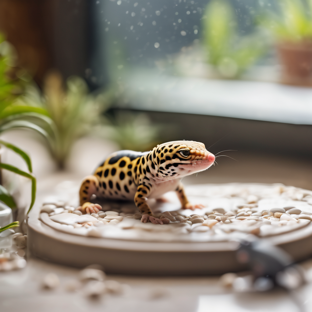
An abscess in a reptile is not a pimple you can pop — it is a walled-off pocket of pus that the animal’s immune system built precisely because it could not drain. Reptiles lack the enzymes that break down pus into liquid, so what forms instead is a firm, caseous lump: dry, cheesy, and semisolid. That single biological fact explains almost everything about how these lumps behave, why squeezing them makes things worse, and why the standard treatment is surgical drainage rather than antibiotics alone.
Abscesses are one of the most commonly reported reptile health problems in practice and one of the most reliably preventable, because the two causes that generate most of them — a bite from live prey and a penetrating substrate injury — are both husbandry choices. This guide covers how abscesses form, where they appear, what you can safely do at home, and exactly when the animal needs a reptile vet.
Quick answer: what a reptile abscess is
An abscess is a localized infection that the immune system has contained. Bacteria — most often from a bite, a scratch, or a retained foreign body — get under the skin, and the body walls them off with fibrous tissue. In a mammal that pocket fills with liquid pus and eventually bursts. In a reptile the inflammatory cells die and dry out instead of liquefying, producing a solid mass of caseous material that has no route out.
Because there is no drainage path, the mass grows until it either erodes through the skin from pressure, becomes large enough to interfere with function, or seeds bacteria into the bloodstream. Antibiotics alone rarely cure an established abscess because the drug cannot penetrate the wall of the capsule in useful concentration. Drainage or surgical removal is the treatment.
The two causes behind most reptile abscesses
1. Live prey bites
This is the number one cause by a wide margin. A cricket, a superworm, a roach or a mouse that is not eaten immediately does not politely wait — it bites, and reptile skin heals by sealing over rather than knitting, so a small puncture closes with bacteria inside it. The classic sites are the ones prey can reach while the animal is not watching: around the eyes, the corners of the mouth, the nostrils, the ear openings, and along the limbs of a lizard resting on the substrate.
Rodent bites are the most dangerous version. A live mouse or rat can inflict deep wounds that seal over and form large abscesses within days, and rodent bites are a well-known cause of the deep, jaw-adjacent abscesses seen in snakes.
2. Substrate and enclosure injuries
Loose particulate substrate abrades and punctures. Sand, gravel, bark chips and wood shavings all cause micro-injuries to the ventrum of a lizard that moves across them, to the rostrum of a snake that pushes through them, and to the skin of any animal burrowing. Those injuries are the entry point. Our substrate guide explains which materials are safe and which cause exactly this class of injury.
Other entry points worth knowing: a shed that went wrong and left retained spectacles or skin tags that became infected, a burn from an unthermostatted heat source, and septicemic spread of infection from elsewhere in the body.
Where abscesses appear, and what that tells you
- Around the eyes and on the head. Almost always a prey bite. Swollen, closed eye with a firm lump behind it is the classic presentation.
- In and around the mouth and jaw. Often confused with infectious stomatitis (mouth rot). The distinguishing feature is that mouth rot is diffuse inflammation of the oral lining, while an abscess is a discrete lump.
- On the limbs and digits. Substrate abrasion, or a constricting shed. These are the most visible and the easiest to catch early.
- On the ventral surface. The signature of a loose substrate problem, particularly in lizards and snakes that rest directly on it.
- Internal. Abscesses in the coelomic cavity or associated with organs present as non-specific illness — weight loss, lethargy, appetite loss — and cannot be diagnosed without imaging. Internal parasite infection and organ abscess produce overlapping pictures.
Signs: how to tell an abscess from everything else
An abscess has a fairly specific feel and behaviour:
- It is firm. Reptile abscesses are hard or doughy, not soft and fluid-filled. Pressing on it does not produce the give you would expect from a fluid-filled lump.
- It is discrete. You can define its edges. Diffuse swelling is usually infection or injury rather than an abscess.
- It grows. Days, not hours, but it does not resolve on its own.
- The animal may not act sick. A single external abscess often leaves appetite and behaviour unchanged. This is exactly why they get missed — the keeper waits for systemic signs that never come until the abscess is large or has burst.
- Eventually the skin over it thins and ruptures, releasing thick, cottage-cheese-like material with a strong odour. If you are seeing that, the abscess has been present for weeks.
The things most often mistaken for an abscess: a retained shed patch (dry, papery, peels), a calcium or fat deposit under the skin (soft, immobile, slow), a hematoma from trauma (bruised, fades over days), a tumor (hard, fixed, slow-growing, non-painful), and a normal fat pad in the tail of a healthy gecko.
Vet warning signs — see a reptile vet promptly:
- Any firm, growing lump anywhere on the body
- An abscess near the eye, in the mouth, or on the head — these are anatomically dangerous and do not wait
- An abscess that has ruptured, is draining, or smells strongly
- Multiple abscesses appearing at once (suggests systemic infection or septicemia)
- Any lump accompanied by lethargy, appetite loss, weight loss, or abnormal posture
- An abscess in a small or juvenile animal, where the mass is proportionally large
Nothing about an abscess improves with waiting. It is a contained infection that either stays contained, gets bigger, or breaks into the bloodstream — and all three outcomes are better handled by a vet than by a hopeful keeper.
What treatment actually involves
Understanding the veterinary approach helps you judge whether a case can wait and what to expect from the visit.
Drainage and debridement
Under sedation or anesthesia, the vet lances the abscess, evacuates the caseous material, and flushes the cavity, usually with an antiseptic solution. In many cases the capsule itself is removed, because a capsule left behind refills. The wound is typically left open to heal from the inside out — closing it would trap the infection again, which is the same mistake the reptile’s own immune system made.
Antibiotics
Systemic antibiotics are prescribed to treat the surrounding tissue infection and prevent spread, but they are the supporting act, not the cure. A reptile with a drained abscess and no drainage is still infected.
Culture and sensitivity
Good practice is to culture the material, because reptile abscesses involve a wide range of organisms — commonly Aeromonas, Pseudomonas, and anaerobes — and the right antibiotic depends on which one. If your vet does not offer this on a significant abscess, it is worth asking about.
Home aftercare
Post-drainage care is mostly keeping the site clean and the animal’s environment immaculate while it heals. Expect a wound that heals slowly by secondary intention over weeks. Keep the enclosure spotless, maintain correct temperatures — reptile healing is temperature-dependent, and a thermostat matters as much during recovery as during health — and complete the full antibiotic course.
What you can safely do at home
Very little, and that is the honest answer. But there are a few genuinely useful things:
- Isolate the animal if it is in a community tank, so cage mates cannot traumatise the lump.
- Switch to frozen-thawed or pre-killed prey immediately. This removes the cause while you arrange the vet visit. Our frozen-thawed feeding guide covers the transition.
- Review the substrate. If it is loose particulate, replace it with solid, non-abrasive flooring. This is both treatment and prevention in one move.
- Keep the enclosure clean and temperatures correct while you wait for the appointment.
- Photograph and measure the lump daily — a coin next to it for scale. This gives the vet an objective growth rate, which is diagnostic information.
What to never do
- Do not squeeze, lance, or drain it yourself. The material is under pressure inside a capsule, and forcing it drives bacteria deeper and into the bloodstream.
- Do not apply human antibiotic ointment to the lump. Reptile skin absorbs topicals readily, and several common ointment bases are toxic to reptiles.
- Do not “flush” it with hydrogen peroxide or alcohol. Both damage reptile tissue severely and delay healing.
- Do not give oral antibiotics from a pet store or a previous prescription. Wrong drug, wrong dose, and it masks the culture result the vet needs.
Prevention: the two habits that eliminate most cases
Never feed live prey
This single change removes the leading cause of reptile abscesses. Frozen-thawed prey, warmed to body temperature, is nutritionally identical, safer for the reptile, and eliminates the bite risk entirely. For the small percentage of animals that refuse frozen-thawed, pre-killed prey — killed immediately before offering — is the fallback. Live feeding in an unattended enclosure overnight is the specific scenario that produces bites and abscesses.
Use non-particulate flooring
Solid substrate — tile, reptile carpet, paper towel, non-adhesive shelf liner — eliminates abrasion injuries to the ventrum and rostrum. If you want a naturalistic setup, use a solid base with a bioactive layer that cannot be ingested in quantity. The drainage-layer approach described in our false-bottom guide gives you the natural look without loose particles underfoot.
The supporting habits
- Never leave an animal on unthermostatted heat. Burns are an entry point. See our burns guide.
- Maintain correct humidity and provide a moist hide so sheds come off cleanly. Retained shed over a toe or spectacle is an infection waiting to happen — our shedding guide covers this.
- Provide proper UVB where the species requires it. Immune function is calcium and vitamin D dependent, and metabolic bone disease weakens every defence the animal has.
- Quarantine and inspect new animals before introducing them.
- Do a weekly hands-on check. Run your fingers over the limbs, the jaw, around the eyes and the vent. Abscesses are found by touch long before they are visible.
Species notes: who is most at risk
- Leopard geckos and other lizards on loose sand — ventral and digital abscesses from substrate abrasion. Leopard gecko care emphasises solid flooring for exactly this reason.
- Snakes fed live rodents — deep bite wounds that seal and form large abscesses, often not noticed until the snake is off food.
- Bearded dragons — crickets left in the tank overnight are the classic cause of eye and facial abscesses. Bearded dragon care covers feeder management.
- Aquatic and semi-aquatic turtles — ear abscesses (aural abscesses) are extremely common and present as a visible swelling behind the tympanum. These need a vet; they do not resolve on their own.
- Chelonians generally — shell injuries that become infected, particularly in outdoor enclosures.
Prognosis: what recovery looks like
A single external abscess, caught before it ruptures, drained properly by a vet and followed by a full antibiotic course, has a good prognosis. Expect the surgical site to take several weeks to close, and expect the animal to behave normally in the meantime.
Abscesses in anatomically awkward places — deep in the jaw, involving the eye, or within the coelomic cavity — are harder and carry a worse prognosis, particularly if function has already been affected.
Multiple simultaneous abscesses, or any abscess with systemic signs (lethargy, appetite loss, weight loss), indicate septicemia, and that is a serious diagnosis with a guarded prognosis even with aggressive treatment. Our emergency warning signs guide lists the systemic indicators to watch for.
Frequently asked questions
Why is my reptile’s lump hard instead of soft and squishy?
Because reptile pus does not liquefy. Reptiles lack the neutrophil enzymes that turn pus into fluid, so the inflammatory material dries into a firm, caseous mass. That is why reptile abscesses feel like a pea under the skin rather than a water balloon, and why they cannot drain on their own.
Can I treat a reptile abscess with antibiotics alone?
Almost never for an established abscess. The fibrous capsule around the pus prevents antibiotics from reaching an effective concentration inside it. Antibiotics treat the surrounding tissue and prevent spread, but the mass itself needs drainage or surgical removal. Very small, early abscesses occasionally resolve with antibiotics, and that is a judgment call for your vet.
My reptile’s abscess burst on its own. Is it fine now?
No. A ruptured abscess means the capsule failed to contain the infection, and material that was under pressure has been released into the enclosure and potentially into the tissue. The animal still needs a vet, the enclosure needs thorough disinfection, and the underlying cause — usually a prey bite or substrate injury — still needs fixing.
Is a lump always an abscess?
No. Fat deposits, hematomas, tumors, retained shed, gouty tophi and normal anatomy all produce lumps. The features that point toward an abscess are firmness, discrete edges, growth over days to weeks, and often a history of live feeding or loose substrate. Anything uncertain goes to a vet — and our transport guide helps you get there safely.
How did my snake get an abscess if I feed frozen-thawed?
Then substrate and enclosure injuries are the likely source, or a wound from an earlier period of live feeding that took weeks to develop. Also check for thermal burns from an unthermostatted heat source and for retained shed. Reviewing the whole enclosure, not just the feeding routine, usually finds the cause.
Will the abscess come back after treatment?
It can, and recurrence usually means one of two things: the capsule was not fully removed, or the cause was not addressed. If your reptile gets a second abscess in a similar location, the answer is almost always still in the husbandry — a substrate that abrades, or prey that can still bite.
Can abscesses spread to my other reptiles?
The bacteria involved can be present in shared water or on shared surfaces, but abscesses themselves are not contagious. The real risk is that the conditions causing them — loose substrate, live prey, poor temperatures — affect every animal in the room. Fix the conditions and you fix the risk for the whole collection.
The bottom line
Reptile abscesses are firm, walled-off pockets of dried pus caused almost entirely by two avoidable things: prey that bites and substrate that abrades. They rarely make the animal look sick until they are large, which means they are found by keepers who touch their animals weekly rather than keepers who wait for behavioural change. There is no effective home treatment — drainage by a vet is the answer — but there is very effective prevention: frozen-thawed prey, solid flooring, and correct temperatures. Change those two habits and abscesses go from the most common problem you deal with to something you read about.
Leave a Reply