
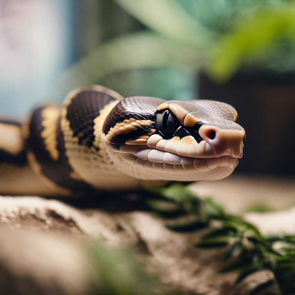
Respiratory infection — keepers call it RI, vets call it URTD or lower respiratory disease — is the single most common reason a pet reptile ends up in a clinic. It is also the most preventable serious illness on this list. A respiratory infection is not a bad-luck pathogen that floats in through the window; it is the end result of weeks of an enclosure that was too dry, too cold, too stagnant, or too dirty, giving an organism that was already sitting harmlessly in the animal’s airway the chance to overgrow. Fix the enclosure and most mild cases resolve on their own. Leave the enclosure alone and the same animal will be back in the clinic within a month.
This guide covers what a reptile respiratory infection actually is, why the pathogen is usually the victim rather than the villain, the early signs that keepers routinely miss, how a reptile vet diagnoses and treats it, the drugs you should not expect to work, and the five husbandry changes that decide whether the infection stays gone. If you keep snakes, geckos, bearded dragons, or any other small pet reptile, this is the health topic most likely to affect you at some point, and the one where what you do in the first 48 hours changes the outcome the most. It belongs alongside our guide to emergency warning signs that need a vet and our species-by-species overview in small pet reptiles: the complete species guide.
What a reptile respiratory infection actually is
A reptile lung is not like a mammal lung. It is a simple, thin-walled sac with very little internal structure — no branching bronchial tree, no diaphragm, and no cough reflex worth the name. Reptiles cannot effectively clear mucus, pus, or fluid from their airways by coughing. That single anatomical fact explains almost everything about how the disease behaves: infections progress slowly, they are quiet, and by the time an animal looks obviously ill the infection has been building for weeks.
Two things happen in an RI. First, the lining of the mouth, trachea, and lung becomes inflamed and produces excess mucus. Second, because the animal cannot clear that mucus, it settles in the lowest part of the lung, where it becomes a growth medium for bacteria. Secondary bacterial infection is what turns a mild irritation into pneumonia. The organisms involved are usually unremarkable — Pseudomonas, Aeromonas, Klebsiella, Mycoplasma, and assorted others that live in soil and water and are present in almost every enclosure on earth. They are opportunistic. They need a reason.
The pathogen is usually the victim, not the villain
This is the part keepers get wrong, and it is why treating the infection without fixing the enclosure fails. Those same bacteria sit harmlessly in the airway of a healthy snake. What lets them overgrow is stress, and in reptiles stress is almost always physical:
- Humidity below the species minimum dries the airway lining, damages the cilia that sweep mucus out, and thins the animal’s first line of defence. Ball pythons in a 40% room will develop RIs that no antibiotic can permanently fix.
- Temperature below the species optimum suppresses immune function directly. A reptile kept at the low end of its gradient runs an immune system at partial power, and respiratory organisms fill the gap.
- Poor ventilation lets humidity and ammonia from waste build up without fresh air exchange. Stagnant, humid, ammonia-tainted air is the classic RI environment.
- Dirty water and substrate raise the bacterial load the animal is breathing and drinking continuously.
- Recent transport, rehousing, or brumation — anything that suppresses the animal’s condition for a week or two.
Antibiotics clear the overgrowth. They do not repair the airway lining, and they do not raise the temperature. Without the husbandry fix, the same bacteria simply regrow. If your reptile has been treated for RI twice, the problem is not the antibiotic — it is the enclosure.
The early signs keepers miss
Early RI is subtle, and the first signs are behavioural rather than respiratory. Run through this list monthly, ideally at feeding time when you are already handling the animal.
Stage one: the behavioural shift
- Mouth held slightly open at rest. A closed mouth at rest is normal. A mouth that sits cracked open when the animal is not thermoregulating or striking is the earliest hard sign, and it is genuinely early.
- Elevated head posture. The animal holds its head higher than usual, sometimes braced against the enclosure wall, to make breathing easier. Snakes in particular will sit with the head raised for long periods.
- Increased time at the warm end that does not resolve after digestion. A reptile running a behavioural fever is trying to raise its body temperature to fight something.
- Loss of appetite out of season, particularly in a species that has been feeding reliably.
- Unusual lethargy. Less exploration, less tongue flicking in snakes, less activity during the active part of the day.
Stage two: the visible signs
- Bubbles at the nostrils, most obvious when the animal is held below the level of its head or after handling. Small clear bubbles are an early sign. Thick white or yellow bubbles are not.
- Clicking, wheezing, or a whistling sound on exhalation. Snakes exhale audibly when the airway is narrowed or wet. Record it on your phone — the vet will want to know whether it is on the inhale or exhale.
- Mucus or discharge from the mouth or nose, progressing from clear to cloudy to opaque.
- Loss of the tongue-flick rhythm in snakes, or flicking that stops early mid-extension.
- Buoyancy or listing in aquatic and semi-aquatic species — an animal that cannot submerge or floats at an angle has fluid in the lungs or coelomic cavity.
Stage three: the signs that mean today, not this week
- Open-mouth breathing at rest, sustained. In reptiles this is a genuine emergency. It is not a dog panting to cool down — it means the animal cannot get enough air.
- Audible gaping with the neck extended and obvious effort in every breath.
- Deep, wet, rattling sounds from deep in the chest.
- Complete refusal to eat for more than two weeks combined with any of the above.
- Any change in colour or perfusion — darkening in a species that pales when warm, or gums and mouth lining that look pale, blue-grey, or injected with red.
Species patterns worth knowing
RI looks slightly different depending on the animal, and knowing your species’ baseline stops both false alarms and missed cases.
- Ball pythons: the classic RI species. Their humidity requirement is 60–70% and a dry room is enough to cause repeated infections. Early sign is almost always a slight open-mouth posture during the day combined with a persistent mid-body head raise.
- Leopard geckos and other dry-housed lizards: RIs are less common but far more dangerous when they happen, because the keeper often does not connect a desert species with a lung infection. Watch for a slightly open mouth, reduced hunting response, and tail fat shrinking faster than the feeding schedule explains.
- Bearded dragons: prone to lower respiratory disease, and the species most often presented too late because their normal open-mouth gaping for thermoregulation masks the real sign. Judge by context: gaping in the morning at the cool end is normal, gaping at 3pm with the animal already warm is not.
- Snakes generally: no cough at all, so mucus simply accumulates. The audible exhale is your single best early indicator, and it is why experienced keepers sit quietly next to the tank at night.
- Turtles and aquatic species: RI presents as pneumonia and often as a swollen, closed, or weeping eye at the same time. Listing and inability to dive is an emergency.
- Amphibians: the picture is different — see our separate guide on amphibian health warning signs, because frogs and newts present skin and balance problems rather than airway mucus.
What your reptile vet will actually do
Exotic vets work from a combination of history and imaging, because a reptile will not cooperate with a stethoscope in any meaningful way. Expect this sequence.
History first — bring the numbers
The vet’s most valuable information is not the exam, it is your enclosure data. Write down and bring: the warm-side and cool-side temperature measured with a digital probe at the animal’s actual resting spot, the day and night humidity, the substrate type, the UVB or heat lamp and its age, the last time the animal fed and what it ate, when the symptoms started, and every treatment you have already tried with doses and dates. A vet who can see that the warm spot has been running at 28 degrees for a ball python has most of the diagnosis before touching the animal. Choosing a vet who actually sees reptiles matters more than almost anything else on this page — read how to choose a vet for your reptile or exotic pet before you need one.
Physical exam
The vet will look into the mouth and glottis for mucus, caseous material, or redness, check for nasal discharge, listen to the lung field through the body wall (limited but not useless in small species), assess body condition by palpation and weight, and check hydration by skin elasticity and eye appearance. In snakes, a cloacal or oral temperature gives a real reading, which is one reason many vets prefer reptiles over small mammals here.
Diagnostics
- Radiographs (X-rays). The standard tool for confirming pneumonia. Reptile lungs sit far forward in the body cavity, and fluid or consolidation shows as increased whiteness in that region. Two views are better than one.
- Tracheal or lung wash. A small amount of sterile saline is introduced and recovered, then cultured and examined under a microscope. This is the gold standard for identifying the organism, and it is the only way to prescribe an antibiotic with any confidence. It requires sedation or anaesthesia.
- Deep nasal or oral swab culture. Easier than a wash and useful, though it can pick up environmental contaminants.
- Blood work. A CBC and chemistry panel to check white cell count, organ function, and hydration. More common in advanced cases and before anaesthesia.
- Faecal exam. Parasites and RI travel together — a heavy parasite load is a common underlying cause of the immune suppression that started everything.
Treatment: what works, what does not
What actually works
Nebulisation is the reptile-specific treatment most keepers have never heard of. Medication is aerosolised into a small sealed chamber and the animal breathes it directly into the lung. It gets drug to the exact tissue that needs it at a fraction of the systemic dose, which matters enormously in a species whose liver and kidney function you cannot easily monitor. Vets commonly nebulise antibiotics, bronchodilators, saline, and occasionally mucolytics. Ask whether your vet can do this or loan you a setup.
Injectable antibiotics are preferred over oral for most reptile RIs. They give reliable absorption regardless of appetite, avoid the gut, and need no cooperation from the animal. Common choices include enrofloxacin, ceftazidime, and amikacin. Your vet will pick based on culture results where available.
Supportive care does more work than owners expect: fluid therapy (subcutaneous or intraosseous in advanced cases), assisted feeding if the animal has not eaten for two weeks or more, and correct warmth throughout treatment. An animal that is warm, hydrated, and eating fights the infection; one that is cold, dry, and anorexic does not, no matter what is in the syringe.
Enclosure correction, which is not optional and not a follow-up item. It starts on day one, before the second injection.
What does not work
- Over-the-counter pet-store antibiotics in the water. Ineffective dosing, wrong spectrum, and it contaminates the water your animal drinks.
- Oral antibiotics when the animal is not eating. Doses get missed, blood levels swing, and resistance develops.
- Steam or a hot shower as a substitute for humidity. Brief exposure does nothing for a chronic dry airway and stresses the animal. Fix the enclosure humidity instead — see reptile humidity control: the complete guide.
- Vitamin C, echinacea, essential oils, or human cold remedies. No evidence in reptiles, and essential oils in particular are actively harmful to species that breathe through skin and airway surfaces directly.
- Cranking the heat to burn it out. Overheating an already compromised animal causes more deaths than the infection.
The husbandry fix that decides whether it comes back
This is the part of the treatment that happens at home, for free, and it is the part that determines the long-term outcome. Five changes, in order of impact.
1. Get the humidity right and hold it
Ball pythons need 60–70%, most tree-dwelling snakes 70–80%, most geckos a dry air with a humid hide, amphibians far higher. Measure it with a calibrated digital hygrometer at the level the animal actually occupies — not a dial on the lid, not a sticker on the glass. Depth of substrate matters more than misting frequency: 3–4 inches of moisture-retaining substrate holds humidity through the day between mistings, while a shallow dry layer swings wildly. Misting once a day into a dry tank spikes the reading for twenty minutes and then returns to 40%. Our guide to measuring temperature and humidity accurately covers calibration and probe placement, and terrarium automation explains how to hold the numbers without standing there with a spray bottle.
2. Fix the gradient, not just the average
An RI animal needs to be able to warm itself on demand. That means a warm side at the species-appropriate top of the range and a cool side, with a thermostat holding the warm spot so it cannot run away. Read why every reptile tank needs a thermostat. During recovery, aim for the upper end of the normal range during the day with a normal night drop — a behavioural fever is the animal’s own treatment and you want to let it happen.
3. Ventilation without desiccation
The classic mistake is solving low humidity by sealing the tank completely, which produces stagnant ammonia-laden air and makes things worse. You need air exchange and humidity at the same time. Reduce the size of the mesh section rather than eliminating it, lower the ventilation but keep it moving, and keep the water dish on the warm side so evaporation feeds the air. Species with high humidity needs in dry climates sometimes need a partially covered top with a humid hide inside rather than a fully sealed box.
4. Clean the environment without sterilising the animal
Spot-clean waste daily, replace water daily, and do a full substrate change at the end of treatment. Do not use strong disinfectants in a tank with a sick animal in it — the fumes and residues matter. Our cleaning schedule for reptile tanks lists which products are safe at which strength. Lower the bacterial load; do not try to eliminate it, because you cannot and the attempt does harm.
5. Remove the stress trigger
Handle as little as possible during recovery — no photoshoot handling, no letting visitors hold the animal. Feed less frequently or smaller prey if the animal is stressed by feeding response, but do not let an underweight animal fast. And re-evaluate anything that changed in the weeks before symptoms started: new substrate, a new lamp, a moved tank, a new pet in the room, a cold snap, a holiday away. That is almost always the trigger. Our first 30 days guide covers the settling period where most first RIs start, and reptile quarantine rules explain why a new animal is the other common source.
Recovery, relapse, and how long to expect
A course of injectable antibiotics typically runs two to four weeks, sometimes longer, and the animal should be improving visibly within the first week. Improving means: mouth closing at rest, head coming down, appetite returning, fewer or no bubbles. If nothing has changed by day seven, tell your vet — the organism may be resistant, the diagnosis may be incomplete, or the husbandry fix has not actually been made.
Expect relapse to be the default outcome if you change nothing. This is the honest number: a reptile treated for RI without a corrected enclosure has a high chance of a second episode within a month or two. A reptile treated with corrected humidity, correct temperatures, better ventilation, and cleaner conditions rarely gets a second one. The antibiotics bought you time; the husbandry bought you the cure.
Weight is your best objective measure. Weigh weekly during recovery and write it down. A snake or gecko that is losing weight while on treatment is not responding, whatever the behaviour suggests. Body condition and weight-based tracking is the same discipline we cover for nutrition in metabolic bone disease in reptiles — slow diseases need slow measurements.
Prevention: the monthly five-minute check
Almost every RI is caught early enough to treat cheaply if someone is actually looking. Once a month, in five minutes:
- Read the hygrometer and the probe thermometer at the animal’s resting spot, and write both numbers down. Compare them to your species’ range — not to what you remember.
- Watch the animal for two full minutes without touching the tank. Is the mouth closed at rest? Is the head held level?
- Listen. Sit quietly for one minute in a silent room. Any click, wheeze, or whistle.
- Check the nose for bubbles, and check under the jaw and along the lower jaw line for any swelling.
- Confirm the last feed date and the animal’s weight trend. Two consecutive weeks of no weight gain in a growing animal, or loss in an adult, is a flag.
Keep a simple notebook or a note on your phone. Trends over six months tell you far more than any single reading, and they are exactly what your vet needs when something does go wrong. Seasonal drift is the most common slow failure — seasonal reptile care explains why your tank’s numbers change with the weather outside and what to do about it.
Frequently asked questions
Can a reptile respiratory infection go away on its own?
Very mild cases sometimes do, if the trigger was a short humidity dip and the enclosure self-corrected. That is not a treatment plan. Because reptiles cannot cough, mucus that settles in the lung does not clear, and what looked like a passing issue often returns as an established infection weeks later. If you can see bubbles or hear a sound, get it checked.
Is reptile respiratory infection contagious to my other reptiles?
The organisms are opportunistic and usually already present in every enclosure, so true animal-to-animal transmission is less common than with mammalian infections. But a sick animal sheds a much higher load, and a stressed or immunologically compromised tank mate can pick it up. Quarantine a symptomatic animal away from others where practical, and never house a recovering animal with a new arrival. Read how long reptile quarantine should be.
My reptile has bubbles from the nose but is eating fine. Do I still need a vet?
Yes. Bubbles mean mucus is being produced faster than it can be cleared, which means the infection is active. Appetite is the last thing to go in reptiles, not the first, and an animal eating normally can still have early pneumonia on X-ray. Early treatment is a short course and a husbandry fix; late treatment is nebulisation, injections, and a real risk of not making it.
Can I treat it at home with antibiotics from the internet?
No. Reptile antibiotic dosing is species-specific and drug-specific, liver and kidney tolerance varies enormously between taxa, and the wrong drug or the wrong dose will either fail silently or cause organ damage you cannot detect until it is advanced. A single vet visit with a culture-directed prescription is cheaper than two failed home attempts and a dead animal.
How do I raise humidity fast for a snake with RI?
Add substrate depth rather than water. Increase to 3–4 inches of moisture-holding substrate, cover most of the mesh top leaving a small vented area, move the water dish to the warm side, and pour water into a corner of the substrate rather than misting the animal. Then measure with a probe at the animal’s level and confirm the reading before you trust it. Do not soak the animal and do not seal the tank completely.
Does UVB help a reptile with a respiratory infection?
Appropriate UVB supports general immune function, calcium metabolism, and appetite, and a species-appropriate UVB setup is good practice regardless. It is not a treatment for RI and it will not substitute for correct humidity, temperature, and antibiotics. Make sure the bulb is the right strength and within its replacement window — see UVB lighting for reptiles.
How do I know my reptile is actually recovering?
Mouth closed at rest, head held at a normal level, no bubbles, no audible breath, steady or rising weight, and normal activity during the active part of the day. All six, not four. The first sign of relapse is usually appetite drifting again combined with a slightly raised head — the same two signs as stage one, which is why the monthly check catches it.
The bottom line
A reptile respiratory infection is a husbandry problem wearing a disease costume. The bacteria are everywhere and harmless to a well-kept animal; what changes is the environment, and then the animal. Treat it early with a vet, support the animal with warmth, fluids, and food, and spend your energy on the enclosure — humidity held through the day, a proper thermal gradient, air that moves, and a tank that is actually clean. Get those five right and this is a one-time problem. Ignore them and it is a monthly appointment. The antibiotics buy time. The setup is the cure.
Leave a Reply